From pixels to prognosis: Imaging redefines liver donor assessment
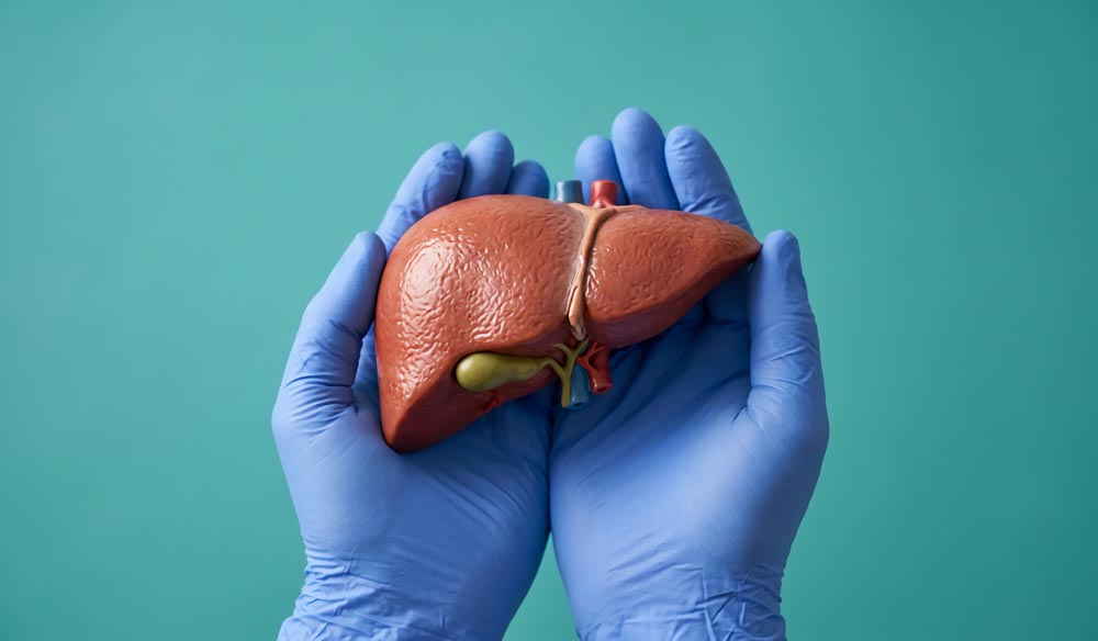
Pre-retrieval imaging is moving liver transplantation toward a more precise and less invasive era. A new evidence synthesis shows how ultrasound, computed tomography, and magnetic resonance imaging can help transplant teams evaluate liver graft quality before retrieval, from detecting fat accumulation to mapping blood vessels, bile ducts, and graft volume. The review also examines how radiomics and artificial intelligence may strengthen decision-making by turning medical images into actionable risk information. By improving graft selection, reducing uncertainty, and supporting better surgical planning, imaging could help transplant teams use more donor organs safely while protecting both donors and recipients.
Liver transplantation often depends on decisions made under pressure. In deceased donation, teams must quickly judge whether a liver is suitable, whether fat accumulation could threaten graft function, and whether unexpected vascular anatomy may complicate procurement or implantation. In living donation, the stakes are different but equally high: surgeons must estimate graft and remnant liver volume accurately and identify biliary variants that could increase the risk of postoperative complications. Biopsy remains important, but it is invasive, time-consuming, and prone to sampling error. Because of these challenges, deeper investigation is needed into how pre-retrieval imaging can improve graft selection, operative planning, and transplant outcomes.
The review was conducted by Andrea Peloso and colleagues from University Hospitals of Geneva, Geneva University Hospital. Accepted (DOI: 10.1016/j.hbpd.2025.12.012) on December 12, 2025, in Hepatobiliary & Pancreatic Diseases International, the article analyzes the role of imaging in deceased and living liver donors. It focuses on steatosis detection, vascular mapping, graft volumetry, and biliary anatomy assessment.
The authors found that each imaging modality offers distinct strengths and limitations. Ultrasound (US) is accessible, inexpensive, and useful as a rapid bedside screening tool, but its accuracy can be reduced by operator dependence, obesity, mild steatosis, and confounding liver changes. Computed tomography (CT) provides fast and objective assessment of hepatic steatosis using attenuation-based measures such as Hounsfield units (HU), liver-to-spleen ratio, and liver-spleen attenuation difference. Its high specificity makes it valuable for identifying grafts that can proceed safely, while borderline cases may still require biopsy. Magnetic resonance imaging (MRI), especially proton density fat fraction (PDFF), offers the most accurate non-invasive quantification of liver fat, although cost, time, and 24-hour availability limit routine use in deceased donors. For vascular assessment, computed tomography angiography (CTA) can map most clinically relevant hepatic arterial and portal venous variants with high agreement to surgical findings. In living donor liver transplantation (LDLT), CT or MRI-based volumetry is generally accurate enough for planning, while magnetic resonance cholangiopancreatography (MRCP) helps identify biliary anatomy but still requires intraoperative confirmation.
The authors said the evidence points to a practical change in how transplant imaging should be understood. Rather than serving only as a preoperative checklist, imaging can become a decision-making framework that links graft quality, surgical feasibility, preservation strategy, and recipient risk. They said future progress will depend on standardized protocols, multicenter validation, and stronger alignment between imaging findings, histology, and intraoperative results. In this model, imaging could become part of a learning health system that continuously improves liver graft assessment.
The review has clear implications for transplant centers working to expand the donor pool without compromising safety. More reliable imaging could help identify usable grafts that might otherwise be declined, while directing higher-risk organs toward biopsy, closer recipient matching, or machine perfusion. For living donors, improved volumetric and biliary mapping can support safer, more individualized surgery. The next step is to validate radiomics and artificial intelligence (AI)-based tools in large, diverse clinical cohorts so they can move from promising research methods to trusted decision-support systems in real-world transplantation.
DOI 10.1016/j.hbpd.2025.12.012
Original Source URL https://doi.org/10.1016/j.hbpd.2025.12.012