
Researchers from UPF and 3D-Shaper Medical take a great leap towards predicting and preventing the risk of hip fracture related to osteoporosis
Researchers at Pompeu Fabra University (UPF) have made a great leap towards predicting the risk of hip fracture among people with osteoporosis and evaluating the effectiveness of specific drugs to prevent it. The BCN Med Tech Unit of the UPF Department of Engineering, together with the company 3D-Shaper Medical, have developed two leading studies in which they present a new computational system to predict and prevent the risk of hip fracture far more precisely than the most common clinical methods today.
With this line of investigation, the researchers are aiming to contribute to preventing and treating diseases like osteoporosis, which has a high social incidence and poses serious health risks. The decrease in bone mineral density, characteristic of osteoporosis, affects the bone’s ability to resist movements or loads. Elderly people of both sexes, but especially women, are most affected by the disease; and, beyond age, cases of secondary osteoporosis can also occur due to medical problems or the intake of certain medication.
Studies to address two key challenges: predicting the risk of hip fracture and the effectiveness of specific drugs to prevent it
Osteoporotic hip fractures generate a high social and economic burden and cause a 20-30% increased risk of mortality, hence their prevention is of the utmost importance. However, the most common diagnostic methods at present -based on dual-energy X-ray absorptiometry (DXA)- do not enable distinguishing between osteoporotic patients who will suffer a fracture and those who will not. Today, assessing drugs’ effectiveness in restoring bones damaged by the disease also remains a challenge.
These are precisely the two challenges addressed by the studies of the BCN MedTech Unit (UPF) and 3D-Shaper Medical. The first, dedicated to predicting the risk of hip fracture with greater precision, has been published in the journal Frontiers in Bioengineering and Biotechnology; and the second, which evaluates the effectiveness of drugs in preventing it, in the Journal of Clinical Densitometry. In both cases, the principal investigators of the studies were Jérôme Noailly (UPF BCN MedTech Unit) and Ludovic Humbert (3D-Shaper Medical).
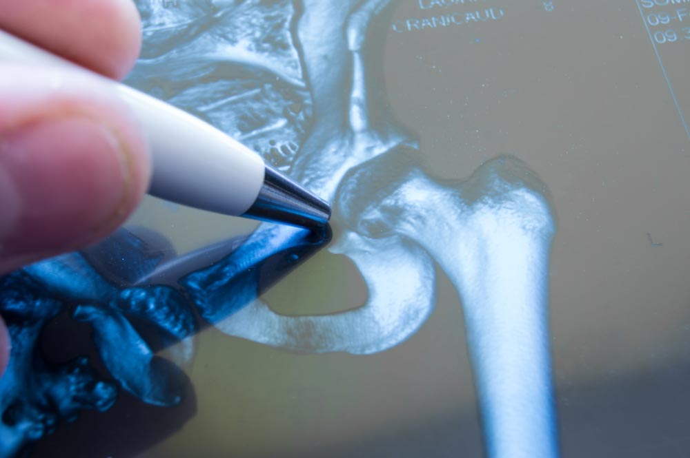
Both studies rely on a new computational system to simulate the functioning of hip bone tissue, based on the reconstruction of 3D images of the joint -using data provided by CETIR Medical Group- and the so-called finite elements (FE) model. It is a very common system in engineering to measure the impact of a movement or mechanical load on the different parts of a structure, in this case hip bone tissue.
The advantages: more accurate diagnoses and treatments and lower clinical costs
Overall, the two studies confirm that the new computational model is a robust alternative to current clinical methods for diagnosing and treating hip osteoporosis. The researchers have shown that computational medicine can assist with the following:
- Refine the stratification of hip fracture risk for osteoporosis: identifying people at higher risk of fracture among those with conditions that make them more vulnerable.
- Optimize drug therapy and treatment: moving towards more precise prescriptions tailored to the needs of each patient.
- Reduce clinical costs related to prevention and treatment: by offering a non-invasive and efficient method to monitor the health status of patients’ hip bones.
The first study simulates stress tests on the hip joint to predict each patient’s risk of fracture
The first study addresses the difficulty in predicting hip fractures among patients with osteoporosis. “Our approach goes beyond bone density, which is what current clinical systems fundamentally measure. We generate a 3D computational model of the patient’s femur -which is part of the hip joint- from a standard 2D image and subject it to a stress test to breaking point”, explains first author Elham Alizadeh, a researcher at BCN MedTech.
This study demonstrates how specific metrics -to measure bone deformation and its resistance to pain under specific loads- contribute significantly to ascertaining which group of patients may suffer a hip fracture and which may not. The new computational system, based on these metrics (or inelastic mechanical descriptors) yields better results than current standard diagnostic systems. It has been successfully tested on a group of 128 women, half of whom had suffered a hip fracture.
The second study creates a digital twin of the hip to evaluate the personalized efficacy of specific drugs
The second study applies so-called digital twin technology to evaluate treatments for osteoporosis. The researchers have simulated the effects of the most common pharmacological treatments on 155 patients with osteoporosis on the bone of the femur -in the part corresponding to the hip joint- over time. In this case, the patients were women and men aged around 40 years who had not received any previous treatment for osteoporosis or whose bone metabolism may have been altered. The computational models have allowed in-depth examination of the mechanical changes induced by these drugs, which has provided precise data on their effects in each patient.
“We have shown that finite element models based on 3D-DXA are sufficiently sensitive to quantify the mechanical benefits of specific drug treatments”, concludes Carlos Ruiz Wills (UPF), first author of this second study, also a researcher at BCN MedTech. This opens the door to major advances in the line of personalized medicine, since this model would allow virtually testing which drug provides the best result for a specific patient before it is prescribed.