Advertisment
Translational science in IBD

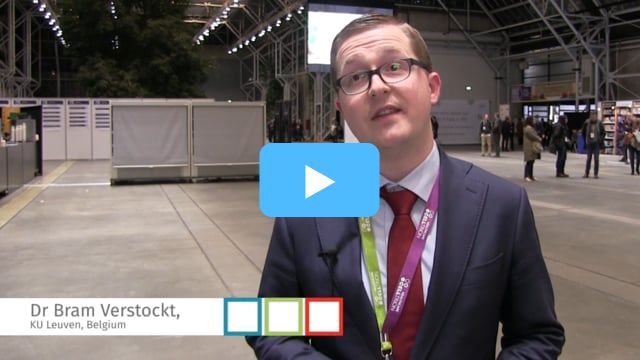
Dr Bram Verstockt (Leuven, Belgium) discusses a biopsy study looking at increases NCR+ ILC3 levels in IBD patients on biologic therapy.
Translational research is playing an increasingly vital role for bridging the gap between, on the one hand, basic science and on the other, clinical studies and routine clinical practice. Among the findings presented in a digital oral presentation session at ECCO 2019 were new insights into the mechanism of action of vedolizumab and potential new therapeutic targets for treating fibrosis and strictures in IBD…
The gut-specific anti-ɑ4β7 integrin antibody vedolizumab has been shown to be highly effective for inducing clinical remission and mucosal healing in UC.1 Dr Marisol Veny (Hospital Clinic Barcelona, Spain) reported the results of a biopsy study to uncover the mechanism of action of vedolizumab and quantify the levels of leukocytes in the intestine and their integrin expression before and after treatment.
Biopsies were collected from a total of eight patients with active UC at the initiation of vedolizumab treatment, and then at 14 and 46 weeks of follow-up. At week 14 two out of seven patients biopsied had achieved remission; at week 46 all eight patients were biopsied, and four were in remission. T cell and B cell levels in the intestine were significantly lower after 14 and 46 weeks of treatment, respectively, compared with baseline. Vedolizumab treatment was also shown to reduce the expression of ɑ4β7 and ɑ4β1 integrins on most lymphocytic populations in blood and intestine.
While vedolizumab-treated patients had increased numbers of circulating ɑEβ7+ memory CD8+ T cells, no effect was seen on the expression of ɑEβ7 integrin in the intestine. The investigators could also show that 100% of ɑ4β7 was bound to vedolizumab in all patients, at all time points in both blood and biopsies, and concluded that vedolizumab works by blocking the migration of T and B cells to the intestinal lamina propria through complete blockade of ɑ4β7 as well as reduced presence of ɑ4β7 on the cell surface (DOP25).
Another biopsy study, reported by Dr Brecht Creyns (University Hospitals Leuven, Belgium), investigated the levels of innate lymphoid cells (ILCs) in blood and biopsies from UC and CD patients treated with either ustekinumab or vedolizumab. This study was based on the rationale that natural cytotoxicity receptor (NCR)-positive ILC3, which produces large amounts of IL-22 in the intestine, has been shown to be decreased in the intestinal mucosa in CD and UC. Colonic biopsies and blood samples were collected from CD and UC patients about to initiate treatment with ustekinumab, vedolizumab or infliximab, and healthy controls at baseline and in connection with endoscopic assessments. The biopsies showed that compared with healthy controls, patients with active disease who were about to begin treatment with vedolizumab or infliximab had significantly lower levels of NCR-positive ILC3 and higher levels of ILC1 at baseline, which recovered by the first endoscopic assessment. In contrast, ILC subgroup levels were normal at baseline in patients about to initiate treatment with ustekinumab. Infliximab and ustekinumab treatment was associated with an increase in peripheral NCR-positive ILC3 levels whereas vedolizumab did not affect NCR-positive ILC3 in the circulation (DOP26).
In a late-breaking abstract, Dr Simonas Juzenas (Christian-Albrechts-University of Kiel, Germany and Lithuanian University of Health Sciences, Kaunas, Lithuania) presented results from a study in which circulating micro-RNA profiles in IBD patients were sequenced to determine their role in IBD aetiology. Micro RNAs are known to be involved in regulating protein-coding gene expression which contributes to a variety of diseases. The investigators sequenced and analysed small RNA transcriptomes from 680 individuals and found evidence of inflammation-specific and disease-specific deregulation of micro RNAs in IBD patients compared with healthy controls. Micro RNAs were identified that correlated with IBD activity and the investigators proposed that these could potentially be used in the diagnosis of IBD (DOP27).
Hypoacetylation of histone-3 lysine 27 (H3K27ac) has been shown to be a pathological feature of stricturing CD. Restoration of histone acetylation using a histone deacetylase (HDAC) inhibitor has been shown to limit fibroblast remodelling and suppress collagen I expression, both key processes in the development of stricturing CD. Dr Amy Lewis (Barts and The London School of Medicine and Dentistry, London, UK) presented transcriptomic data on genes that were up- or down-regulated in human colon fibroblast cells cultured with valproic acid, which acts as an HDAC inhibitor. Valproic acid was found to reverse the effects of the TGF-β1 cytokine including decreased H3K27 acetylation, increased phosphorylated SMAD3 levels, and increased cytoplasmic collagen I protein and secreted pro-collagen-IαI (DOP20).
Another study which focused on fibrosis in IBD was presented by Dr Bruce Weder (University of Zurich, Switzerland) and was based on the rationale that IBD patients with active disease have decreased luminal intestinal pH, and that extra-cellular acidification is known to promote fibroblast activation and extracellular matrix remodelling. The investigators obtained human fibrotic and non-fibrotic terminal ileum from CD patients undergoing ileocaecal resection due to stenosis at centres in Switzerland and the Netherlands and analysed these for markers of fibrosis and expression of the pH-sensing G protein-coupled receptor 4 (GPR4). GPR4 expression was found to correlate significantly with the expression of pro-fibrotic cytokines and increased collagen deposition. In a mouse model, mice that lacked GPR4 expression had decreased collagen layer thickness and less expression of fibrosis marker micro RNA (DOP24).
Based on presentations at the 14th Congress of ECCO, 6–9 March 2019, Copenhagen, Denmark.
References
1. Feagan BG, Rutgeerts P, Sands BE, et al. Vedolizumab as induction and maintenance therapy for ulcerative colitis. N Engl J Med 2013;369:699-710.


