Advertisment
Melanoma by smartphone apps?

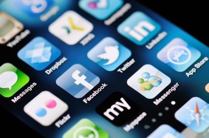
Smartphone applications that claim to evaluate a user’s photographs of skin lesions for the likelihood of cancer instead returned highly variable and often inaccurate feedback, according to a study led by researchers at the University of Pittsburgh School of Medicine. The findings, published in JAMA Dermatology and available online today, suggest that relying on these “apps” instead of consulting with a physician may delay the diagnosis of melanoma and timely, life-saving treatment.
“Smartphone usage is rapidly increasing, and the applications available to consumers have moved beyond communication and entertainment to everything under the sun, including health care,” said lead researcher Laura Ferris, M.D., Ph.D., assistant professor, Department of Dermatology, University of Pittsburgh School of Medicine. “These tools may help patients be more mindful about their health care and improve communication between themselves and their physicians, but it’s important that users don’t allow their ‘apps’ to take the place of medical advice and physician diagnosis.”
In fact, the study found that three out of the four smartphone applications tested incorrectly diagnosed 30 percent or more melanomas as “unconcerning” based on their evaluation of user images.
The study, funded by the National Institutes of Health (NIH) and the University of Pittsburgh Clinical and Translational Science Institute, reviewed applications available in the two most popular smartphone platforms and found that such tools often are marketed to nonclinical users to help them decide, using a digital image for analysis, whether or not their skin lesions are potential melanomas or otherwise concerning, or if they likely are benign. Researchers uploaded 188 images of skin lesions to each of the four applications, which then analyzed the images in different ways, including automated algorithms and images reviewed by an anonymous board-certified dermatologist. The applications often are available for free or at a very low cost, and are not subject to any regulatory oversight or validation.
Only the application that utilized dermatologists for a personal review of user images, essentially functioning as a tool to facilitate teledermatology, provided a high degree of sensitivity in diagnosis – just one of the 53 melanomas was diagnosed as “benign” by the experts reading the images. This application also was the most expensive, costing users $5 per image evaluation. Although the tools included disclaimers stating they were providing information for educational purposes only, researchers noted the risk that patients might rely on the application’s evaluation rather than seek the advice of a medical professional.
The likelihood of relying on the application’s free or low-cost evaluation is particularly concerning for the uninsured or economically disadvantaged, especially because a substantial number of melanomas are first detected by patients, noted the study authors. “If they see a concerning lesion but the smartphone app incorrectly judges it to be benign, they may not follow up with a physician,” said Dr. Ferris. “Technologies that decrease the mortality rate by improving self- and early-detection of melanomas would be a welcome addition to dermatology. But we have to make sure patients aren’t being harmed by tools that deliver inaccurate results.”
###
Co-authors of the study include Oleg Akilov, M.D., Timothy Patton, D.O., Joseph C. English III, M.D., Jonhan Ho, M.D., Joel A. Wolf, and Jacqueline Moreau, all of the University of Pittsburgh.
The study was funded by NIH grants UL1RR024153 and UL1TR000005.


