Less-invasive cardiac MRI is a valuable diagnostic tool in the early evaluation of patients with acute chest pain
An estimated 3 million patients visit emergency departments each year with acute chest pain and mildly elevated troponin levels. High levels of troponin, a protein, occur when the heart muscle is damaged from a heart attack. How best to evaluate and treat patients with chest pain with detectable or mildly elevated troponin remains unclear.
Now, a new study from researchers at Wake Forest University School of Medicine reveals that cardiac magnetic resonance imaging (MRI), is a safe and valuable tool to help evaluate these complex patients.
The study findings appear online today in Circulation: Cardiovascular Imaging, a journal of the American Heart Association.
“Patients who present to the emergency room with chest pain and mildly elevated troponin often fall into a diagnostic gray zone,” said Chad Miller, M.D., professor and chair of emergency medicine at Wake Forest University School of Medicine and emergency medicine physician at Atrium Health Wake Forest Baptist. “It’s not readily clear whether they should have angiography or other forms of testing when being evaluated in the emergency department.”
Angiography is used to check the health of blood vessels and how blood flows through them, but angiography procedures are more invasive than MRI. For example, during cardiac catheterization, a catheter is guided through a patient’s blood vessel to the heart. Providers use the test to identify conditions such as clogged arteries.
For the study, researchers randomized 312 participants at four sites in the U.S. to either cardiac MRI or more invasive-based interventions with modification as needed when conditions evolved. The sites included William Beaumont Hospital in Royal Oak, Mich., the University of Mississippi in Jackson; The Ohio State University Wexner Medical Center in Columbus; and Atrium Health Wake Forest Baptist Medical Center in Winston-Salem.
Participants were followed for 2.8 years. The average age of study participants was 61; 60% were men, 64% were white, and 34% were Black. All participants experienced acute chest pain and troponin levels between detectable and 1.0 ng/ml.
The research team compiled data on heart attacks, deaths, and cardiac-related hospital readmission or emergency visits.
“We did not detect any differences in clinical or safety event rates between cardiac MRI and the invasive-based care pathway,” Miller said. “We also found that using cardiac MRI reduced the need for invasive angiography over the long-term follow-up period.”
In the cardiac MRI group, 58% of participants were safely discharged based on negative imaging and did not have angiography or an intervention such as revascularization within 90 days. Revascularization involves treatment to restore blood flow to a section of the heart that is blocked.
“These findings confirm that cardiac MRI is a highly accurate test that can be reliably used as first-line testing in this complex patient population,” Miller said.
Support from the study was provided by the National Heart, Lung and Blood Institute grant No. R01HL118263
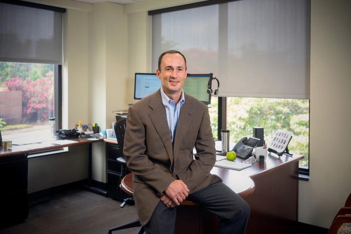
IMAGE: CHAD MILLER, M.D., PROFESSOR AND CHAIR OF EMERGENCY MEDICINE AT WAKE FOREST UNIVERSITY SCHOOL OF MEDICINE AND EMERGENCY MEDICINE PHYSICIAN AT ATRIUM HEALTH WAKE FOREST BAPTIST. view more
CREDIT: WAKE FOREST UNIVERSITY SCHOOL OF MEDICINE